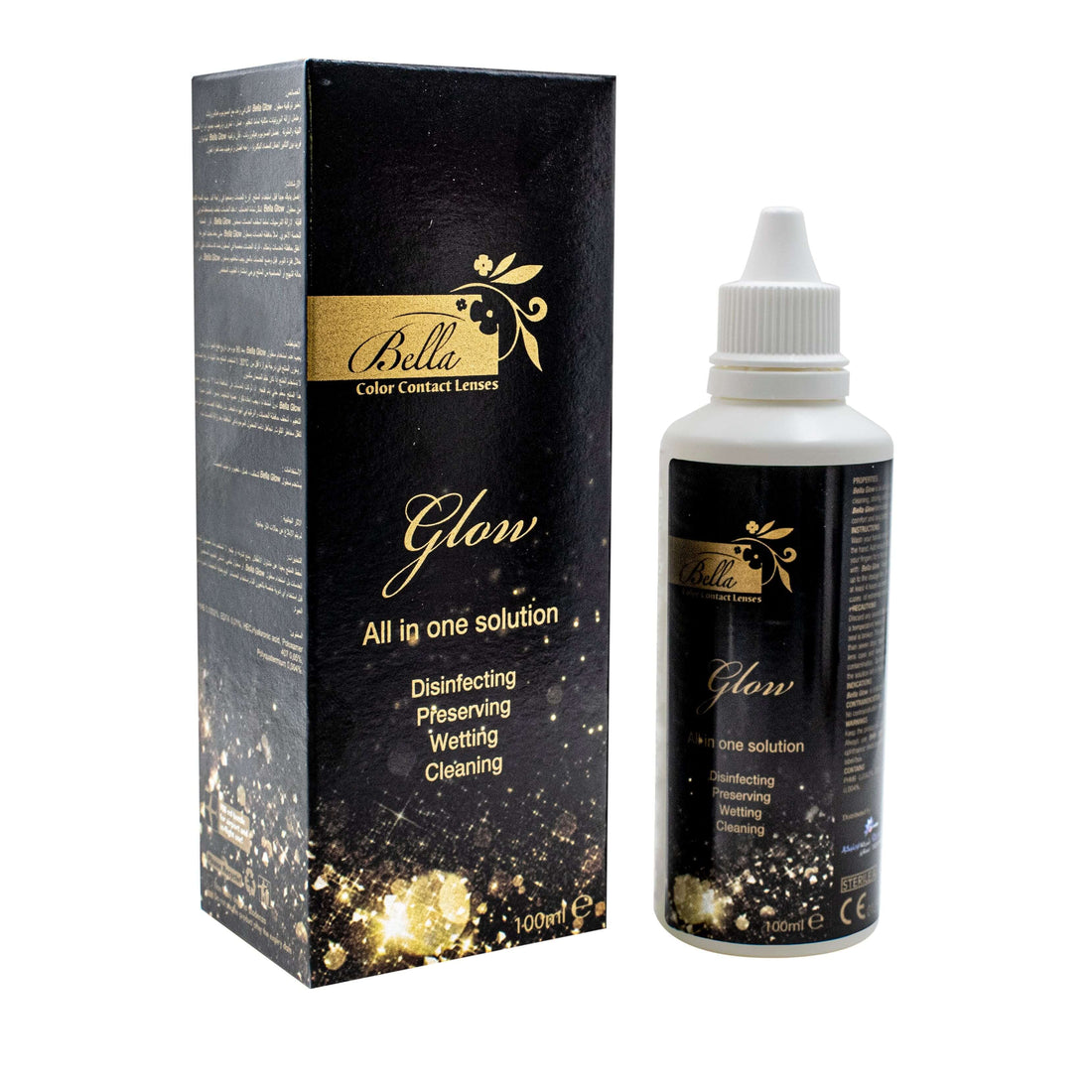

Contact lens keratitis is a corneal infection that can develop quickly and, without treatment, cause lasting damage to your vision. If you have removed your lenses and are experiencing pain that is not easing, significant redness, or sensitivity to light, understanding what type of infection may be involved and what to do next is the most important step you can take right now.
Quick Answer
Contact lens keratitis is an infection or inflammation of the cornea caused or aggravated by contact lens wear. It presents in four forms: bacterial, fungal, viral and Acanthamoeba. Symptoms include eye pain, redness, light sensitivity and blurred vision. It requires same-day professional assessment and cannot be safely managed at home.

What Is Contact Lens Keratitis?
Contact lens keratitis refers to infection or inflammation of the cornea, the clear tissue at the front of the eye, in people who wear contact lenses. The cornea is the eye's first line of defence, but contact lenses change its surface environment in ways that make infection more likely. In the UK, corneal infection affects around four in every 10,000 contact lens wearers each year, according to data from Moorfields Eye Hospital. It is one of the most serious complications of contact lens wear and requires prompt medical attention.
How Contact Lenses Create Conditions for Infection
A healthy cornea has natural defences against infection: the tear film flushes microorganisms away, and the epithelial surface acts as a physical barrier. Contact lenses disrupt both mechanisms. They hold bacteria, fungi, and parasites against the corneal surface rather than allowing tears to wash them away. Small breaks in the epithelium, which can occur without any obvious injury, provide an entry point for microorganisms that would otherwise pose little risk. Wearing lenses beyond recommended schedules, using tap water near lenses, or sleeping in them significantly weakens these natural protections.
How Quickly Does Contact Lens Keratitis Develop?
The speed of development depends on the type of infection. Bacterial contact lens keratitis, the most common form, can progress from initial exposure to significant corneal damage within 24 to 48 hours. Acanthamoeba keratitis, by contrast, often has a delayed presentation with symptoms appearing days to weeks after exposure, which is one reason it is frequently misdiagnosed in its early stages. The rapid progression of bacterial keratitis means that any worsening eye pain after lens removal should be assessed the same day rather than monitored overnight.
The Four Types of Contact Lens Keratitis
Not all cases of contact lens keratitis are caused by the same organism, and the type matters significantly for both urgency and treatment. The four forms differ in how quickly they develop, how serious they become, and which contact lens habits place wearers at greatest risk. Understanding the differences helps in recognising warning signs specific to each type.
Four Types of Contact Lens Keratitis
|
Type |
Caused by |
Onset speed |
Severity |
Highest risk factor |
|---|---|---|---|---|
|
Bacterial |
Pseudomonas aeruginosa, Staphylococcus aureus |
Hours |
High |
Overnight lens wear, poor case hygiene, skin bacteria transfer |
|
Acanthamoeba |
Waterborne parasite (Acanthamoeba spp.) |
Days to weeks |
Very high |
Water contact with lenses, tap water use for rinsing or storage |
|
Fungal |
Candida, Fusarium, Aspergillus |
Days |
High |
Tap water storage, exposure to soil or plant material |
|
Viral (HSV) |
Herpes simplex virus |
Hours to days |
Moderate to high |
Pre-existing HSV infection, immune stress, corneal trauma |
Bacterial keratitis accounts for approximately 90 per cent of microbial keratitis cases in contact lens wearers and is the type most likely to develop from everyday hygiene lapses. Acanthamoeba keratitis is less common but disproportionately severe, and the UK has a higher incidence than many countries due to how domestic water is stored and distributed.

Bacterial Keratitis: the Most Common Type
Bacterial contact lens keratitis most commonly involves Pseudomonas aeruginosa, a bacterium that adheres readily to soft contact lens surfaces, or Staphylococcus aureus, which is found naturally on skin and can be transferred to lenses during handling. The infection typically causes intense, rapidly worsening pain, significant redness, and a visible white or grey spot on the cornea representing an ulcer. Because bacterial keratitis can progress to corneal perforation if left untreated, it is treated as a medical emergency even when the initial presentation appears moderate.
Acanthamoeba Keratitis: Why It Is Particularly Serious for UK Wearers
Acanthamoeba is a microscopic organism found in tap water, rivers, lakes, swimming pools and hot tubs. Moorfields Eye Hospital has noted that the UK has a higher incidence of Acanthamoeba keratitis than many other countries, linked in part to how domestic water is stored. The infection is uniquely difficult to treat because Acanthamoeba can form a hardy cyst stage that resists standard disinfectants and even dedicated anti-amoebic medications. Treatment typically requires months of intensive topical therapy, and in some cases corneal transplantation is necessary. A distinctive feature of Acanthamoeba keratitis is pain that feels far more severe than the visible redness would suggest. This is an important clinical warning sign.
Fungal and Viral Keratitis
Fungal contact lens keratitis, most commonly caused by Fusarium or Candida, develops more slowly than bacterial keratitis and often results from contact with organic matter such as soil or plant material. Using tap water to rinse or store lenses also raises the risk significantly. Treatment involves antifungal eye drops and, in some cases, oral antifungal medication sustained over several weeks. Viral keratitis caused by herpes simplex virus tends to present with dendritic lesions on the corneal surface and is treated with antiviral drops or tablets. It can recur and, with repeated episodes, may cause permanent corneal scarring that affects vision.
Recognising the Symptoms of Contact Lens Keratitis
The symptoms of contact lens keratitis overlap with those of less serious lens-related conditions such as overwear syndrome or dry eye. The difference is usually in the intensity, the direction of symptoms after lens removal, and whether certain specific features are present. Recognising which pattern applies to your situation determines whether you have hours or minutes to act.
Symptom Severity and Response Guide
|
Symptom pattern |
Likely interpretation |
What to do |
|---|---|---|
|
Mild redness and grittiness improving within 2 hours of lens removal |
More likely overwear or dry eye than contact lens keratitis |
Remove lenses; rest; contact optometrist if symptoms are not resolving |
|
Redness, burning and light sensitivity persisting or worsening after lens removal |
Possible early contact lens keratitis |
Same-day optometrist assessment; do not reinsert lenses |
|
Intense pain, discharge, blurred vision, white or grey spot on cornea |
Active contact lens keratitis |
Go to A&E or NHS eye casualty today; bring lens case and solution |
|
Severe pain out of proportion to redness, ring-pattern infiltrate visible, recent water exposure |
Possible Acanthamoeba keratitis |
A&E immediately; tell clinicians about water contact and lens use |
The key distinction between overwear discomfort and contact lens keratitis is that keratitis symptoms typically worsen rather than improve after lens removal. Any eye pain that intensifies overnight or is accompanied by visible changes to the corneal surface should be treated as keratitis until professionally assessed.

The White Spot on the Cornea: What It Means
A visible white or grey spot on the cornea is one of the most reliable visible signs of active contact lens keratitis. It represents a corneal ulcer, meaning an area where the corneal epithelium has broken down and infection has taken hold in the underlying tissue. The spot may be small initially, but bacterial ulcers can enlarge rapidly over hours. If you can see a white area over the coloured part of your eye when looking in a mirror, or if someone else notices it, this requires same-day assessment at A&E or eye casualty. Do not wait for a GP referral.
Why Acanthamoeba Keratitis Pain Feels Disproportionate
Acanthamoeba keratitis is characterised by a level of pain that appears far greater than the visible signs in the eye. Many patients describe intense, continuous aching or burning while the eye appears only moderately red. This pattern of pain out of proportion to clinical appearance is well documented in the literature and is a specific warning sign for Acanthamoeba infection. If you have had water exposure while wearing lenses and are experiencing this pattern, mention the water contact to clinicians when you arrive at A&E, as it changes the management approach significantly.
Symptoms That Distinguish Contact Lens Keratitis from Overwear Syndrome
Both overwear syndrome and contact lens keratitis can cause redness, grittiness, and discomfort after a long day of lens wear. The distinguishing features of contact lens keratitis are symptoms that worsen after lens removal rather than improving, pain that is sharp or aching rather than a dull gritty sensation, visible changes to the corneal surface, and discharge that is thick or purulent rather than watery. Our article on contact lens infection symptoms covers the full range of signs that distinguish different types of lens-related conditions.
When to Seek Help: A Clear Triage Guide
The most common error people make with contact lens keratitis is waiting to see how things feel in the morning. Unlike overwear discomfort, keratitis does not resolve with rest alone. Delays in treatment, particularly for bacterial or Acanthamoeba forms, can result in permanent corneal scarring that is not reversible. The framework below describes where to go based on the pattern of symptoms you are experiencing.
Triage: Where to Go and How Quickly
|
Level |
Symptoms |
Where to go |
How quickly |
|---|---|---|---|
|
Routine monitoring |
Mild redness improving after lens removal, no persistent pain |
Optometrist at next scheduled aftercare |
Next routine appointment |
|
Urgent: same day |
Redness not improving after 2 hours, any persistent eye pain, early light sensitivity |
Call optometrist practice; request same-day urgent slot |
Within hours; same day |
|
A&E or Eye Casualty |
Intense pain, vision changes, visible white spot, worsening after lens removal, overnight deterioration |
NHS Eye Casualty or A&E; no referral needed |
Go immediately |
|
A&E immediately |
Severe pain + water exposure history + ring-pattern discolouration of cornea |
A&E directly; tell staff about contact lens use and water contact on arrival |
Do not delay for any reason |
NHS Eye Casualty departments at major eye hospitals across the UK accept walk-in patients and do not require a GP referral. If you cannot reach your optometrist and symptoms fall in the urgent or emergency categories, going directly to the nearest eye casualty is the appropriate course of action.
How Is Contact Lens Keratitis Treated?
Treatment for contact lens keratitis depends on the type of organism involved, which is why the infecting pathogen must be identified, usually through corneal scrape and culture, before the correct medication can be prescribed. All contact lens wear must stop immediately upon diagnosis and should not resume until a clinician has confirmed the infection has fully resolved and the cornea has healed.

Bacterial Keratitis Treatment
Bacterial contact lens keratitis is treated with intensive antibiotic eye drops, typically fluoroquinolones such as ofloxacin or ciprofloxacin, applied very frequently in the initial stages, often every one to two hours including through the night for the first 24 to 48 hours. The frequency reduces as the infection responds to treatment. Most patients require follow-up appointments every one to two days in the early treatment period. Severe cases with deep corneal involvement may require fortified antibiotic drops prepared by hospital pharmacies and, in some instances, short-term admission.
Acanthamoeba Keratitis Treatment
Acanthamoeba keratitis requires a different and considerably more demanding treatment protocol. A combination of antiseptic agents, typically chlorhexidine and propamidine, is applied very frequently and maintained for weeks to months. Treatment is continued long after symptoms improve because Acanthamoeba in its cyst form can persist for extended periods and cause relapse if therapy ends prematurely. Some patients require surgical intervention, including corneal transplantation, if significant scarring has occurred. Early diagnosis is critical because outcomes are substantially better when treatment begins before the infection reaches the deeper corneal layers.
What to Expect During Treatment
Throughout treatment, contact lens wear is stopped completely. Most patients rely on glasses for the entire treatment period and for some weeks beyond. Follow-up appointments are frequent and non-negotiable; missing them can allow the infection to become resistant or spread to deeper tissue. Preservative-free lubricating drops may be used for comfort, but over-the-counter redness-relief drops that contain vasoconstrictors should be avoided during treatment, as they can mask signs of progression and give a false impression of improvement.
Recovery Timeline for Contact Lens Keratitis
Recovery from contact lens keratitis varies considerably depending on the type of infection, how early treatment began, and whether the infection reached the deeper layers of the cornea. Early presentation and prompt treatment are the strongest predictors of a full recovery with minimal lasting impact on vision. The figures below represent typical ranges.
Recovery Timeline by Severity
|
Scenario |
Typical recovery time |
Outcome if untreated |
|---|---|---|
|
Mild bacterial keratitis, treatment within 24 hours |
1 to 2 weeks with antibiotic drops and close follow-up |
Likely full visual recovery |
|
Moderate bacterial or fungal keratitis |
3 to 6 weeks; possible change of medication |
Some residual corneal scarring possible |
|
Severe bacterial keratitis with ulceration |
6 to 12 weeks; possible specialist referral |
Corneal scarring; reduced vision in affected area |
|
Acanthamoeba keratitis |
3 to 12 months of intensive treatment |
Possible corneal transplantation; some permanent visual loss in severe cases |
Individual recovery depends heavily on the specific organism, the corneal layers affected, and how promptly treatment began. Your treating clinician will provide a more specific prognosis based on your examination findings and how the infection responds to initial therapy.

When Can You Return to Wearing Contact Lenses After Keratitis?
Returning to contact lens wear after contact lens keratitis should only happen once a clinician has confirmed the infection has fully resolved: the epithelial surface is healed and there are no active signs of infection. This is typically a minimum of several weeks after treatment ends for mild cases, and potentially considerably longer for moderate or severe episodes. When you do return, your optometrist will likely recommend a lens type with higher oxygen permeability and a wearing schedule that reduces your risk of recurrence.
Contact Lens Keratitis Risk Factors and Coloured Lenses
Understanding which behaviours and circumstances increase the risk of contact lens keratitis is the most direct path to prevention. Some risk factors apply to all contact lens wearers. Others are particularly relevant to coloured lens users and are rarely addressed in depth, despite being an important part of responsible coloured lens use in the UK.
Behaviours That Significantly Increase Keratitis Risk
The behaviours most strongly associated with contact lens keratitis are sleeping in lenses not approved for overnight wear, which raises infection risk sixfold or more according to published clinical research; exposing lenses to any water source including tap water, shower water, swimming pools or hot tubs; wearing lenses beyond their replacement date; and failing to clean and replace the lens case regularly. Our guide to showering with contact lenses explains the specific risks of water exposure in detail, including what to do if your lenses have already come into contact with water.
Unregulated Coloured Contact Lenses and Keratitis: the UK-Specific Risk
In the UK, all contact lenses including plano coloured lenses are classified as medical devices under MHRA regulations and must carry a CE mark. A study published in PubMed found that a significant number of cosmetic contact lens websites selling to UK consumers do not meet the requirements of the Opticians Act, and that many lenses sold through those channels lack CE marking.
Lenses without CE marking may be manufactured without sterile processes, may contain pigments that are not fully encapsulated, or may use materials that do not maintain adequate oxygen permeability. All of these factors directly increase the risk of contact lens keratitis.
Our guide to buying coloured contact lenses safely covers how to verify that a retailer meets UK requirements before purchasing any coloured lens.
Why Regulated Coloured Lenses Carry a Different Risk Profile
Regulated coloured contact lenses from UK-registered retailers are manufactured to the same safety standards as clear medical lenses. The pigment is fully encapsulated within the lens material so it does not contact the ocular surface, oxygen permeability is maintained to clinical specifications, and sterile packaging is verified.
The risk of contact lens keratitis associated with quality regulated lenses worn within recommended schedules is not meaningfully higher than for equivalent clear lenses, provided hygiene practices are followed consistently. Checking expiry dates on lens packaging is also part of responsible lens use; our article on whether contact lenses expire explains why expired lenses raise infection risk even before they cause noticeable discomfort.
Bella Lense sells only CE-marked coloured contact lenses through a UK-registered platform, in compliance with MHRA medical device regulations. If you want to wear coloured lenses with confidence in their safety, browse our coloured contact lens collection to find options that meet UK safety standards and are backed by proper optometrist fitting guidance.

Preventing Contact Lens Keratitis
The majority of contact lens keratitis cases are preventable. The behaviours that cause most preventable cases are well established, and changing them consistently is the most effective protection available. The following habits address the highest-risk behaviours across all contact lens types, including coloured lenses.
- Never sleep in lenses not specifically approved for overnight wear, including during short naps.
- Keep all water away from your lenses and lens case, including tap water, shower water, swimming pools and hot tubs.
- Replace your lens case every month, allow it to air dry after each use and never rinse it with tap water.
- Wash and thoroughly dry hands before handling lenses; bacteria from skin are the primary source of bacterial keratitis.
- Follow the replacement schedule for your lens type exactly, even if the lenses still feel comfortable and appear undamaged.
- Purchase coloured contact lenses only from UK-registered retailers selling CE-marked lenses, and attend an optometrist fitting before wearing any new lens type.
Frequently Asked Questions
What is contact lens keratitis?
Contact lens keratitis is an infection of the cornea linked to contact lens wear. It occurs in four forms: bacterial, Acanthamoeba, fungal and viral, and requires professional treatment.
Can contact lens keratitis cause blindness?
In severe or untreated cases, yes. Bacterial and Acanthamoeba keratitis can cause corneal scarring and permanent vision loss. Early treatment significantly reduces this risk.
How quickly does keratitis develop from contact lens wear?
Bacterial keratitis can develop within 24 to 48 hours. Acanthamoeba keratitis has a delayed onset of days to weeks, making early recognition particularly important.
How long does treatment for contact lens keratitis take?
Mild bacterial cases typically resolve in one to two weeks. Acanthamoeba keratitis can require three to twelve months of intensive treatment in more serious cases.
Can I wear coloured contact lenses safely without getting keratitis?
Yes, when lenses are CE-marked, professionally fitted, worn within recommended schedules, and cared for properly. The risk relates to behaviour and lens source, not colour itself.
What is the white spot on my eye after wearing contacts?
A white spot on the cornea is a corneal ulcer, one of the signs of active keratitis. It requires same-day assessment at A&E or an NHS eye casualty unit.
How do I know if I have keratitis or just lens irritation?
Keratitis symptoms worsen after lens removal rather than improving. Key signs include pain that intensifies overnight, visible corneal changes, and discharge thicker than normal watering.
Conclusion
Contact lens keratitis is one of the most serious complications of lens wear, but it is both preventable and treatable when caught early. The decisions you make in the first few hours after noticing severe symptoms have a direct impact on outcomes. If pain is not improving after lens removal, same-day professional assessment is not an overreaction. It is the right call.
Medical Disclaimer
The information in this article is intended for general educational purposes only and does not constitute medical advice. If you are experiencing symptoms of contact lens keratitis or any ocular pain or infection, remove your contact lenses immediately and seek same-day assessment from a registered optometrist, A&E department, or NHS eye casualty unit. Do not delay care or attempt to self-treat based on this content.